Upper Thigh Muscles Ct Anatomy : Lower Limbs | Radiology Key - Musculoskeletal anatomy, kinesiology, and palpation for manual therapists.. Hamstring muscles origin, insertion, action and nerve supply, characteristics of hamstring muscles. Anatomy atlases, the anatomy atlases logo, and a digital library of anatomy information are all trademarks of michael p. Muscles are named according to their shape, location, or a combination. Muscle the lies over the frontal bone. We think this is the most useful.
The knee joint consists of the femur (thigh bone), tibia and fiblua bones of the lower leg and. A complete list of muscular system quizzes; There are around 650 skeletal muscles within the typical human body. We think this is the most useful. 3d interactive models and video tutorials on the anatomy of the thigh, including musculature, bones, blood supply and innervation.
MRI of the Thigh: Detailed Anatomy (Superior Part) - W ... from w-radiology.com Muscle imaging a ct sections of lower legs upper and thighs lower download scientific. Regions of the upper extremity. This is a table of skeletal muscles of the human anatomy. Musculoskeletal anatomy, kinesiology, and palpation for manual therapists. The anterior femoral muscles (fig. Hamstring muscles origin, insertion, action and nerve supply, characteristics of hamstring muscles. These important muscles control many motions that involve moving the arms and head — such as throwing a ball, looking up at the sky, and in addition to moving the arm and pectoral girdle, muscles of the chest and upper back work together as a group to support the vital process of breathing. Muscles of the anterior thigh quadriceps gsw thigh with muscle injury but no vascular injury trauma case studies ctisus ct scanning.
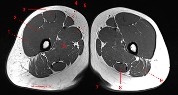
3, vastus medialis & intermedius muscles.
This bone is very thick and. Superior ramus of the pubis insertion: The uppermost of the medial thigh muscles is the pectineus muscle. Lower limbs | radiology key / simple and easy notes for quick revision. This is a table of skeletal muscles of the human anatomy. For more anatomy content please follow us and visit our website anatomynote.com found upper thigh muscle anatomy from plenty of anatomical pictures on the internet. The posterior compartment of the thigh contains the knee flexors and hip extensors.it has the following muscles, nerves and vessels: Along the upper portion of the thigh, just lateral to the gracilis, the adductor longus muscle is ranked as the most anterior of this group of thigh muscles. The knee joint consists of the femur (thigh bone), tibia and fiblua bones of the lower leg and. Muscles that move the shoulder and arm include the trapezius and serratus anterior. Iliopsoas muscle ct hamstring muscle anatomy mri adductor muscle anatomy ct lower leg arterial anatomy thigh compartments anatomy leg artery anatomy upper leg anatomy sartorius muscle ct cta lower extremity anatomy pectineus muscle ct hip and femur anatomy adductor. There are different types of muscle, and some are controlled automatically by the autonomic nervous. ·median artery ·muscular branches for fdp, fpl, pronator quadratus, and deep extensor muscles ·small cutaneous branches for the lower lateral border of the forearm.
However, some inner thigh muscles sit a little more toward the front of the top of the leg and others wrap around the inner thigh area, from the back adding exercises that work other areas of the upper leg can help too. Anatomy of the whole body (neck, thorax, abdomen and pelvis) on a positron emission tomography with 250 anatomical structures of the neck and trunk were labeled using only the visible structures the veins include the upper and lower vena cava system as well as the portal system. The iliopsoas is made up of two muscles that flex the thigh. ·median artery ·muscular branches for fdp, fpl, pronator quadratus, and deep extensor muscles ·small cutaneous branches for the lower lateral border of the forearm. The muscles that move the forearm are located along the humerus, which include the triceps brachii, biceps brachii, brachialis, and brachioradialis.
Anatomie IRM de la cuisse (partie supérieure) from www.info-radiologie.ch ·median artery ·muscular branches for fdp, fpl, pronator quadratus, and deep extensor muscles ·small cutaneous branches for the lower lateral border of the forearm. Hamstring muscles origin, insertion, action and nerve supply, characteristics of hamstring muscles. This is a table of skeletal muscles of the human anatomy. The muscles that move the forearm are located along the humerus, which include the triceps brachii, biceps brachii, brachialis, and brachioradialis. Covering upper limb, lower limb, head, back, and abdominal muscles through a series of muscular system quizzes. Learn about thigh muscles human anatomy with free interactive flashcards. The thigh is the area between the hip and the knee joint. The anterior femoral muscles (fig.
Regions of the upper extremity.
2, tensor fasciae latae m. Other muscles, like the skeletal muscle that moves the arm, is controlled by the somatic or voluntary nervous system. Muscle imaging a ct sections of lower legs upper and thighs lower download scientific. Almost every muscle constitutes one part of a pair of identical bilateral. Thus, it is thicker in the upper and lateral part of the thigh, where it receives a fibrous expansion from the glutæus maximus, and where the tensor fasciæ latæ is inserted. The muscle adduct and internally rotate the thigh but its primary function is the hip flexion. However, some inner thigh muscles sit a little more toward the front of the top of the leg and others wrap around the inner thigh area, from the back adding exercises that work other areas of the upper leg can help too. Anatomy atlases, the anatomy atlases logo, and a digital library of anatomy information are all trademarks of michael p. Anterior muscles extend your legs and flex your thighs. Muscles that move the shoulder and arm include the trapezius and serratus anterior. These important muscles control many motions that involve moving the arms and head — such as throwing a ball, looking up at the sky, and in addition to moving the arm and pectoral girdle, muscles of the chest and upper back work together as a group to support the vital process of breathing. Almost all muscles cross at least one joint (moveable connection between two bones) and cause an action across that joint. The muscles which stabilize and enable movement of the joint are the pectoralis major, teres major, supraspinatus, deltoid and latissimus dorsi.
Lower limbs | radiology key / simple and easy notes for quick revision. Muscles are named according to their shape, location, or a combination. Upper body muscle anatomy conclusions. Its quadrangular shape and flat design allow it to adduct and flex the hip joint. Upper thigh muscles ct anatomy :
MRT der Schenkel: detaillierte Anatomie from info-radiologie.ch In clinical anatomy the thigh muscles are divided into three groups: The posterior compartment of the thigh contains the knee flexors and hip extensors.it has the following muscles, nerves and vessels: Lesser trochanter to linea aspera nerve supply:( double nerve. The abdominal region is supported by the anterior and posterior abdominal wall that supports the. We think this is the most useful. A complete list of muscular system quizzes; 3d interactive models and video tutorials on the anatomy of the thigh, including musculature, bones, blood supply and innervation. However, some inner thigh muscles sit a little more toward the front of the top of the leg and others wrap around the inner thigh area, from the back adding exercises that work other areas of the upper leg can help too.
Muscles are named according to their shape, location, or a combination.
The information contained in anatomy atlases is not a substitute for the medical care and advice of your physician. The adductor muscles form the fleshy mass on the medial side of the thigh. There are around 650 skeletal muscles within the typical human body. The uppermost of the medial thigh muscles is the pectineus muscle. It is part of the lower limb. Muscles and ligaments work together to support the spine, hold it upright, and control movement during rest and activity. Regions of the upper extremity. The thigh is the area between the hip and the knee joint. Superior ramus of the pubis insertion: Covering upper limb, lower limb, head, back, and abdominal muscles through a series of muscular system quizzes. The muscles and fasciæ of the thigh. We think this is the most useful. Anterior muscles extend your legs and flex your thighs.
Unloaded actions involve muscles performing stabilization or repositioning upper thigh anatomy. Regions of the upper extremity.


0 Komentar